
Seeing Clearly: Conservation of Wax Eye Moulages
CONTENT WARNING
- diseased and injured human eyes -
CONTENT WARNING - diseased and injured human eyes -
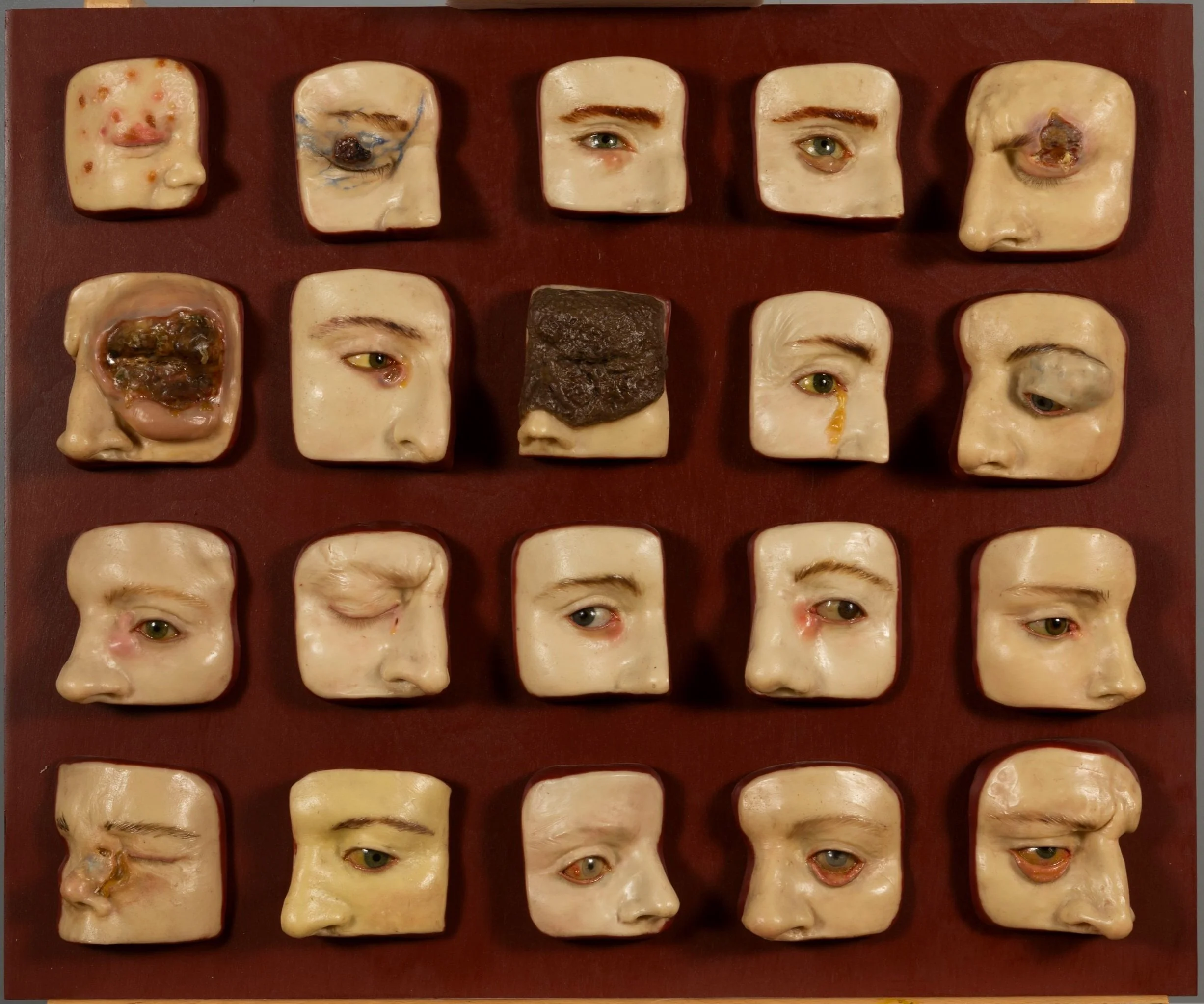
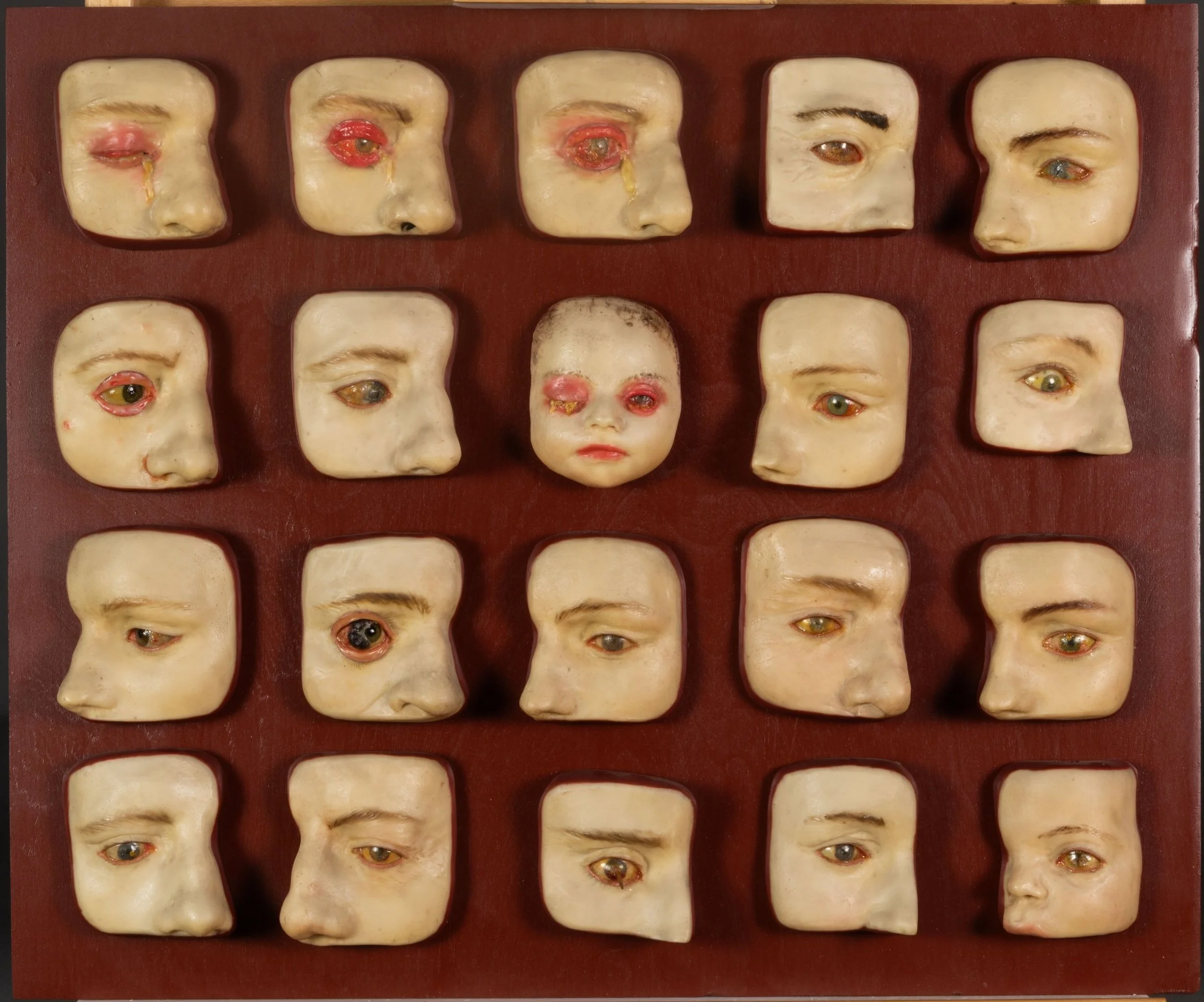
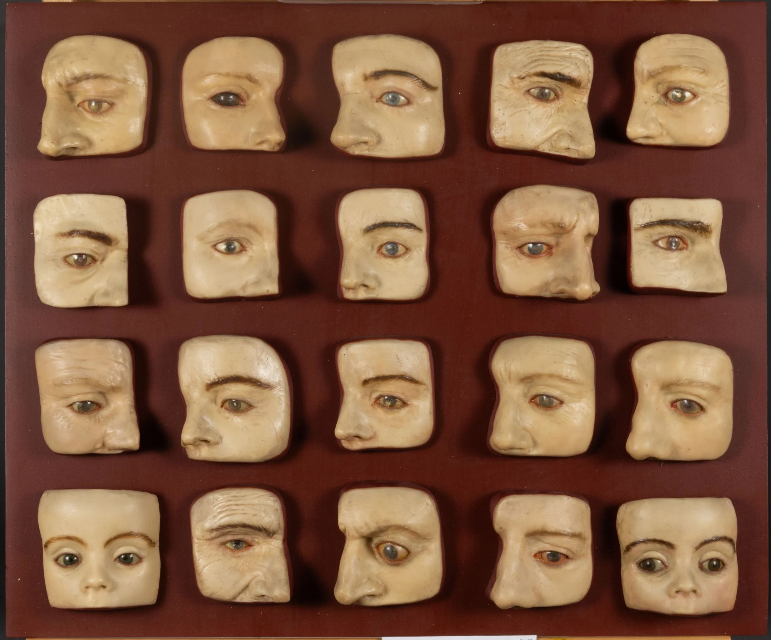
In 2025, Flux staff had the opportunity to treat a set of 100 anatomical wax eye moulages from the Mütter Museum, a medical history museum in Philadelphia, PA. The moulages were mounted on five separate panels, and being over 150 years old were now in varying states of condition, as some were on display for years, some had been restored previously and one set had been kept in storage. As an iconic visitor favorite, the eye moulages were in need of thorough cleaning, cornea consolidation and replacement, loss compensation of missing eyelashes, and retouching of previously mismatched repairs.
Brief History of Anatomical Wax Models
Renewed interest in the study of anatomy during the Renaissance led to the creation of a new wax art form: wax preparatory models. From as far back as ancient Greece, the study of anatomy was limited by both social and practical reasons. Historically, it was considered morally and socially unacceptable to dissect a human body, and in many instances even illegal for a long time. By the time of the Renaissance, there were means to legally obtain a cadaver for dissection and scientific study, but the number of legal cadavers available was not enough to meet the demand. Renaissance anatomists usually had to resort to illegal methods, such as grave robbing. There was also the unpleasant task of having to dissect and examine unpreserved dead bodies, which necessity required to be confined to the winter months or a very strong stomach. With anatomical studies becoming more scientifically-based, access to more readily available materials for teaching purposes became necessary. And so, in response to this need, artists began to create wax anatomical models, and these soon became preferred by anatomists for their ability to surpass the limitations of two-dimensional drawings when a cadaver was not available.
The Mütter Museum moulages were created in the 19th century by the Vasseur-Tramond workshop in Paris. The workshop was operated by Pierre Vasseur and his son-in-law Gustave Tramond. Most workshops viewed their techniques and recipes as trade secrets, Vasseur-Tramond included, and as a result there is limited information about the specifics of the process or materials used. We examined the moulages using 365 nm ultraviolet radiation (UV-A) and a stereomicroscope to get a closer look.
Materials
After examining the moulages and conducting research we made some interesting observations. In one of the primary reference publications, the authors note that “in the great majority of Tramond’s pieces the technique employed involved the use of wax ‘velaturas’; that is, on the plaster mould of the desired figure fine translucent layers of a kind of wax known as ‘Izmir wax’, coloured with natural pigments dissolved in animal oils, were laid on top of each other. The filiform structures, such as vessels and nerves, were produced by bunches of thread impregnated in colored wax...A common practice in most countries was the use of organic items such as human teeth and hair.”
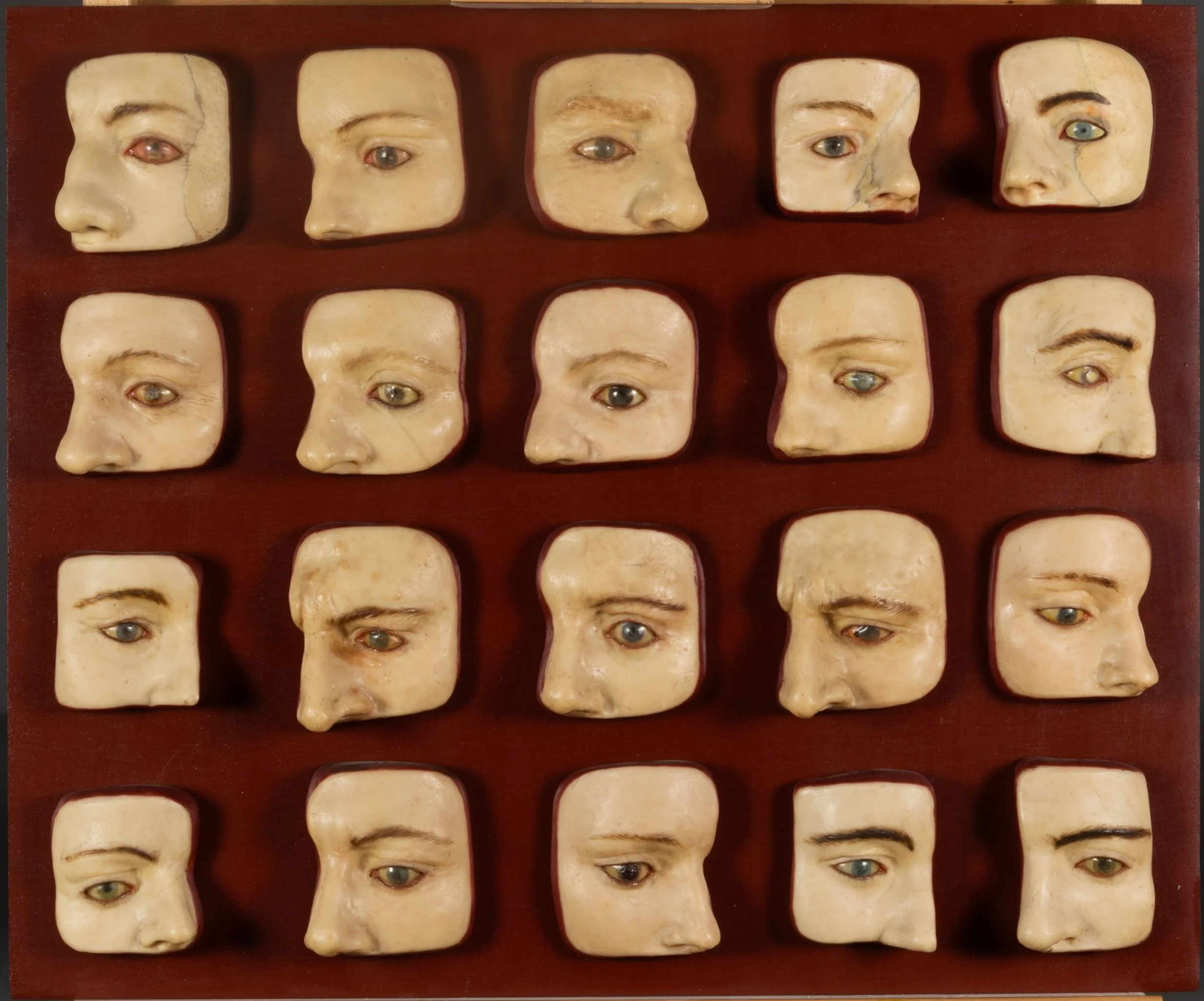
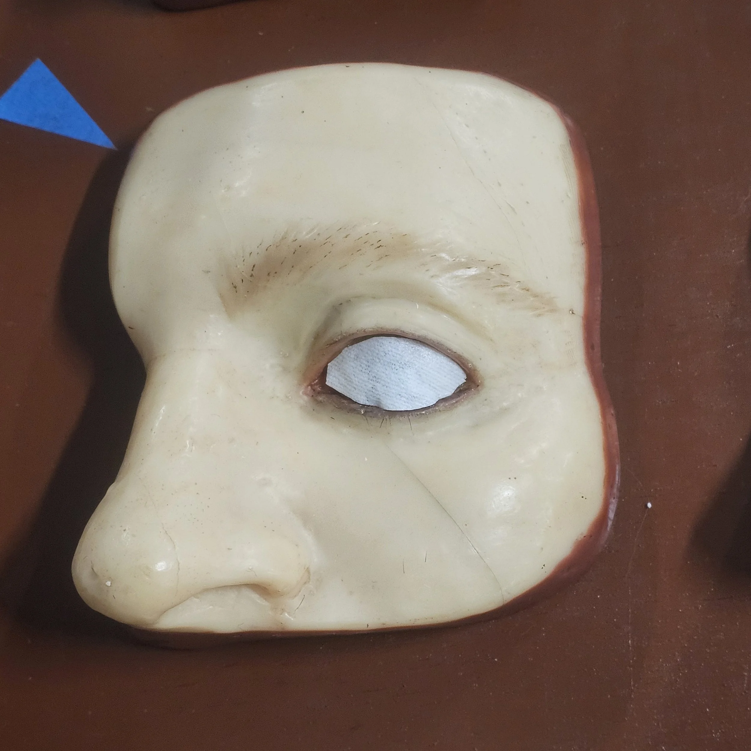
This description aligns with our observation of the group of Mütter eye moulages, particularly the likely use of repeated plaster molds to create the faces upon which the various eye pathologies were depicted. Material characterization through archival research and published instrumental analysis shows in general the moulages comprise beeswax that has been modified and made more elastic through the addition of a variety of materials including turpentine, natural resins, animal fats and various pigments, applied in thin layers in order to achieve the desired consistency and coloration.
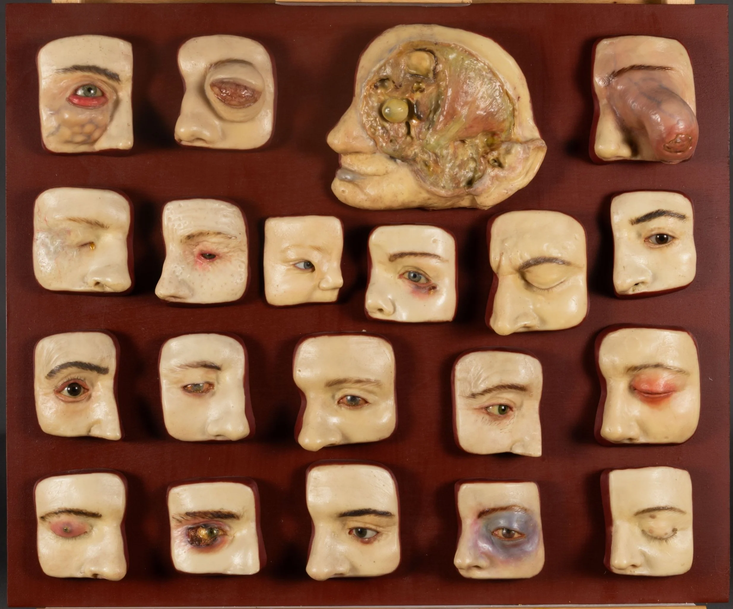
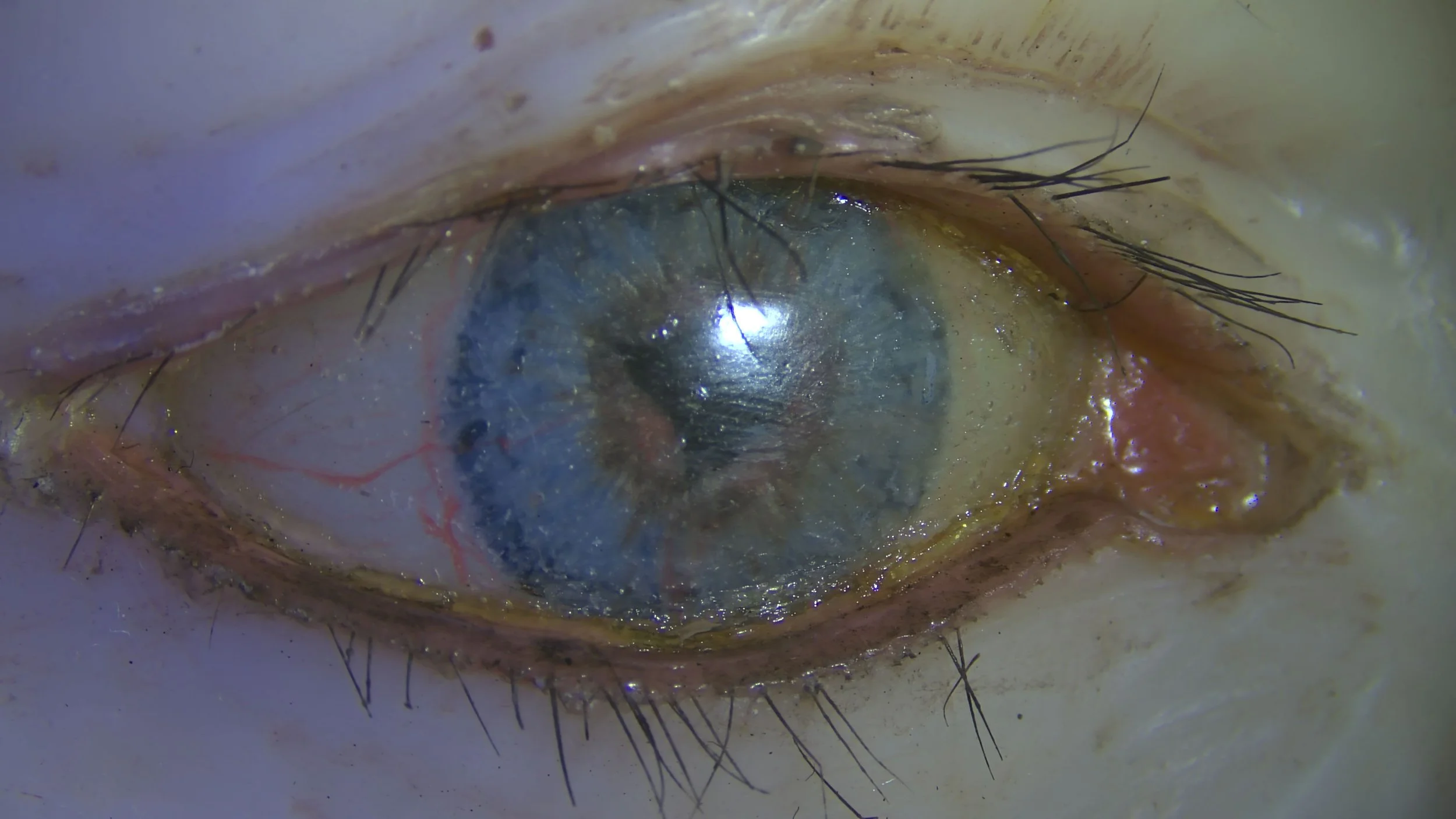
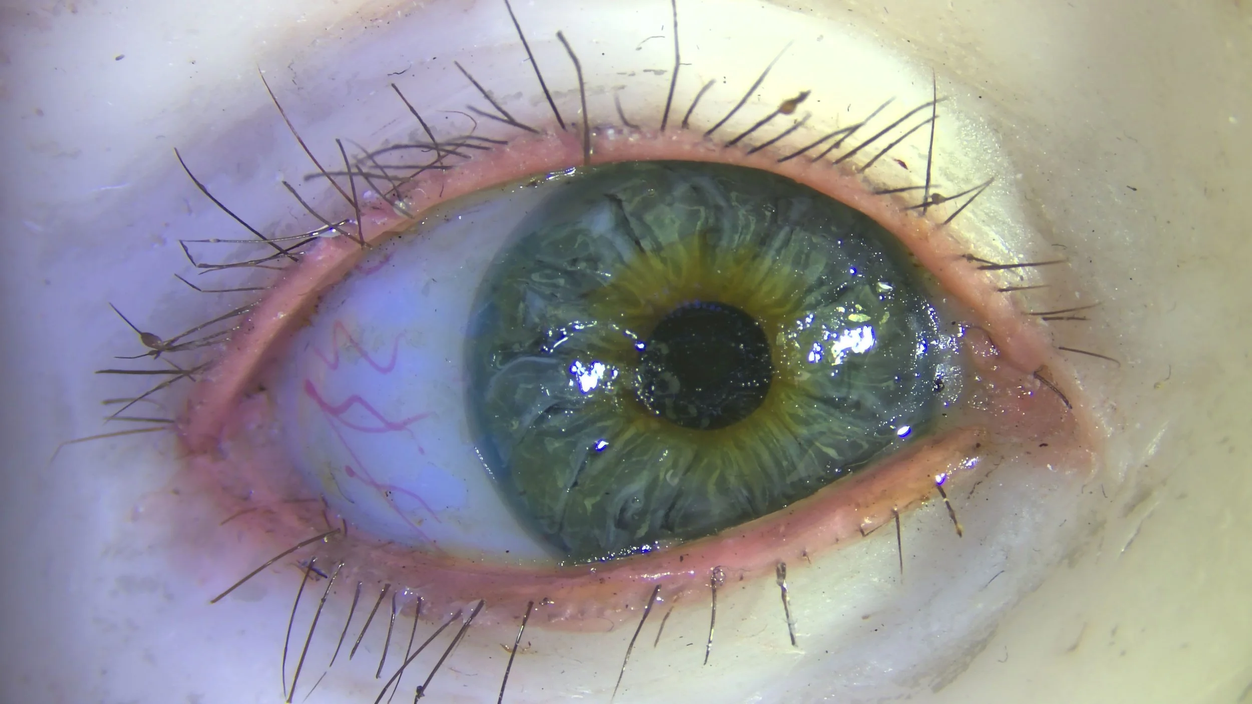
Closer examination of the eyes – the cornea, sclera, and iris - using the stereomicroscope revealed at least 2 types of eyes: reverse painted glass eyes and wax eyes. Both types of eyes were further enhanced with coatings, paint and pigmented wax to create the individual pathologies. Capillaries are depicted using dyed threads as well as painted lines, and sometimes both.
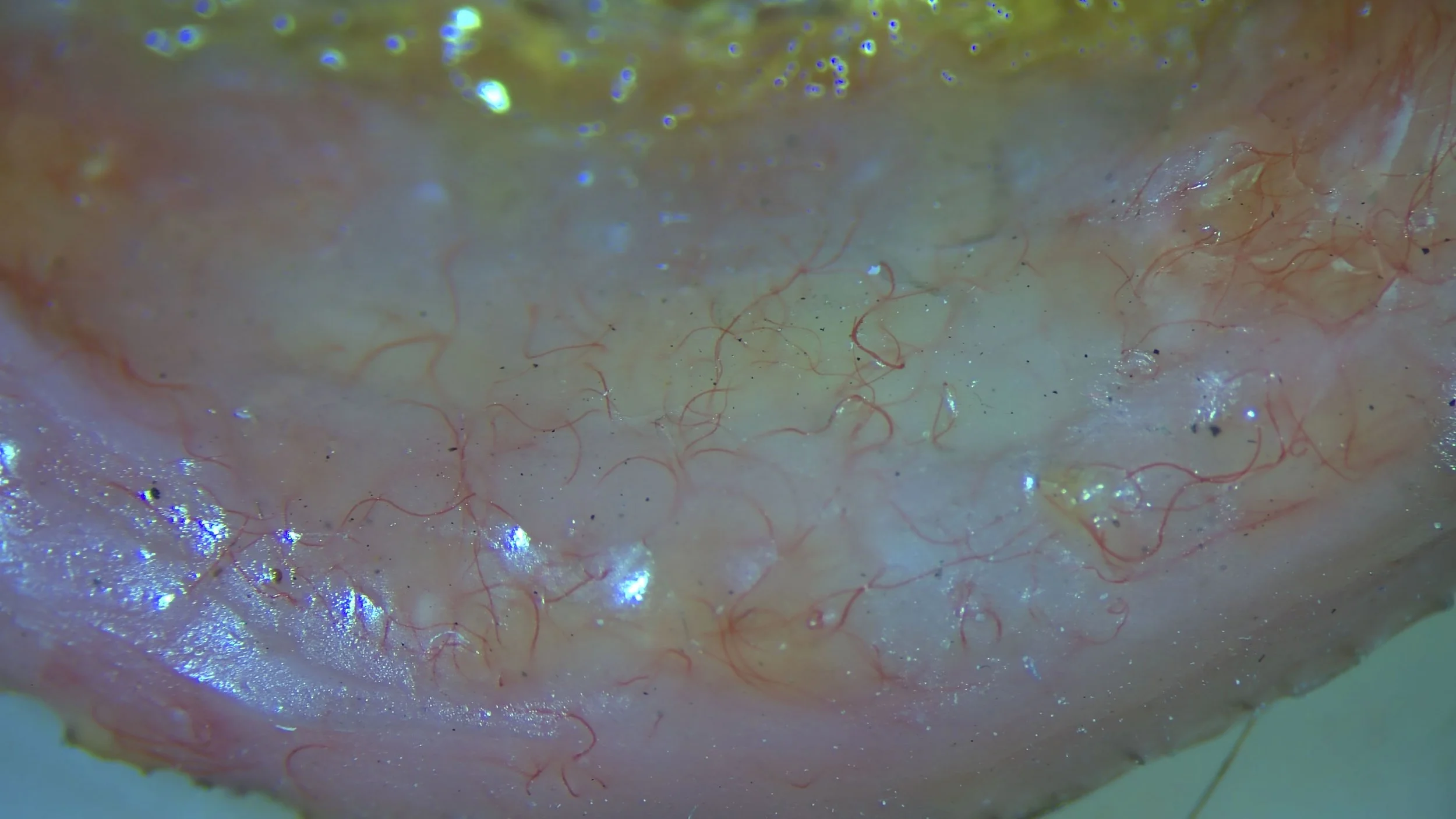
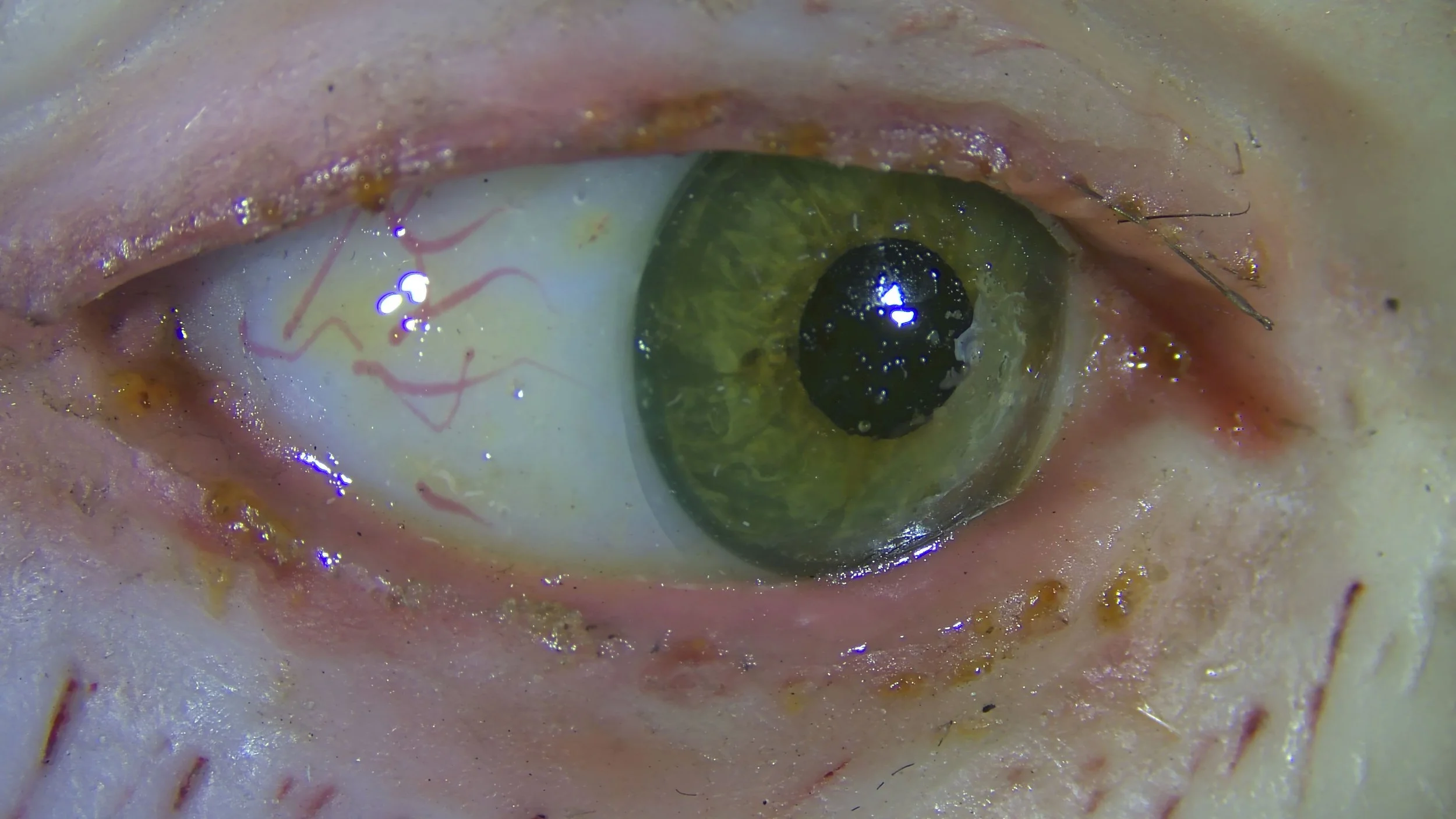

Fig. 4 details, before treatment, UV
The crimson pigment
Based on its characteristic orange-pink fluorescence when viewed under UV - as seen in the picture - and its historical use in wax models, the crimson color used to replicate the rosy cheeks and inflamed skin on these wax eye moulages is likely cochineal.
The cochineal is a cactus-eating insect historically used to create brightly colored cochineal dye. It takes about 70,000 insects to make one pound of cochineal dye. Cochineal extract can be further purified to create pigments in shades of pink, red and purple. These natural dyes have been used historically in cosmetics, food coloring and paint and textiles.
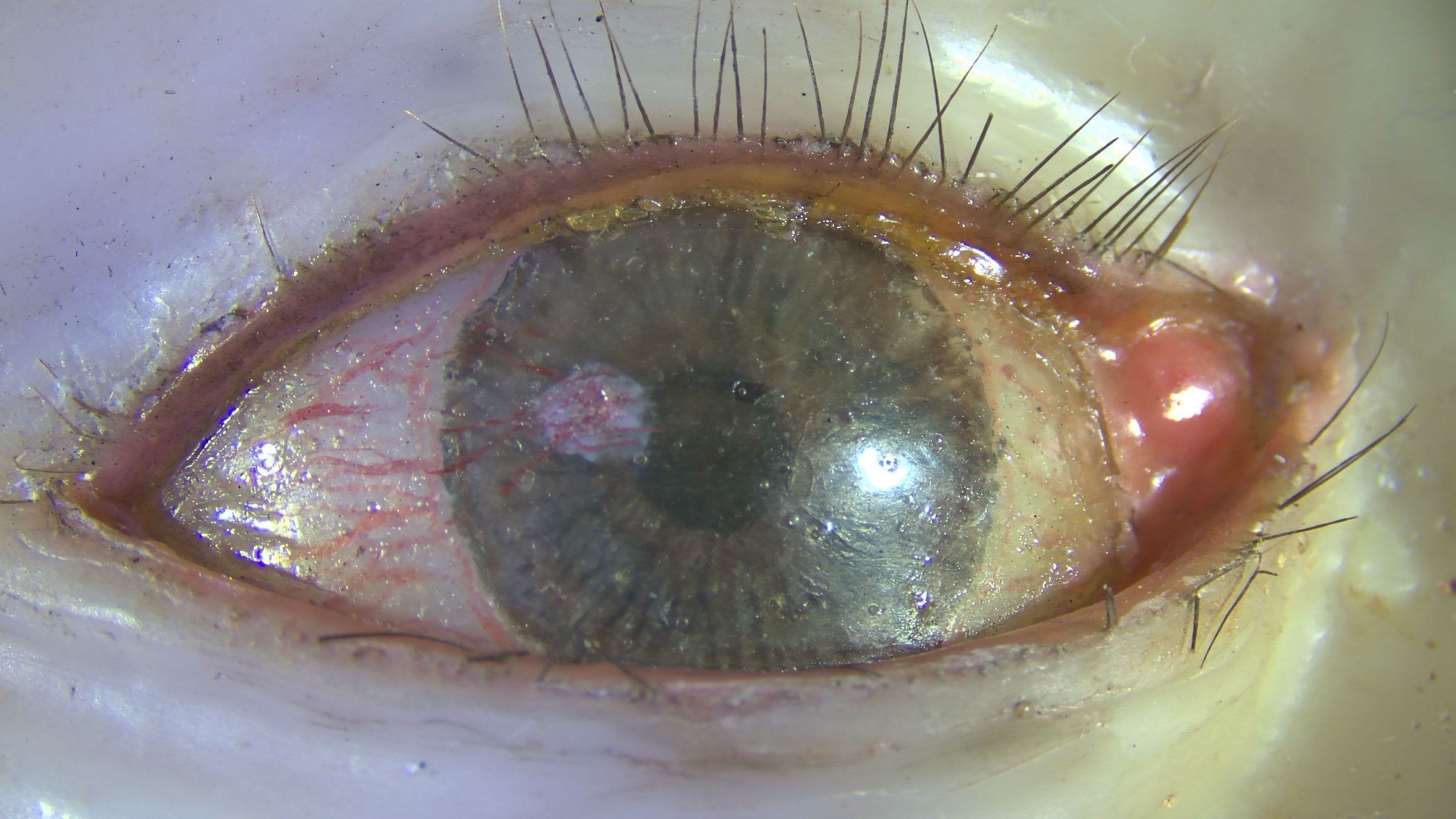
The eyelashes
Research done on a Vasseur-Tramond life-size wax anatomy model of a female's head and chest at the University of Valladolid Anatomy Museum found that the hair on their model was human and that natural bone, specifically a skull, jaw and teeth, were used as internal supports.
Based on this information, it is reasonable to believe that the eyelashes on the Mütter Museums’ wax eye models are also human eyelashes or the cut ends of human hair.
Cleaning the wax
We can tell from the visible retouching and fills that the eyes have received restoration treatment before, though we don't know when or by whom. Some moulages also exhibit microscopic loss of pigment and surface details, suggesting aggressive cleaning solutions have been used.
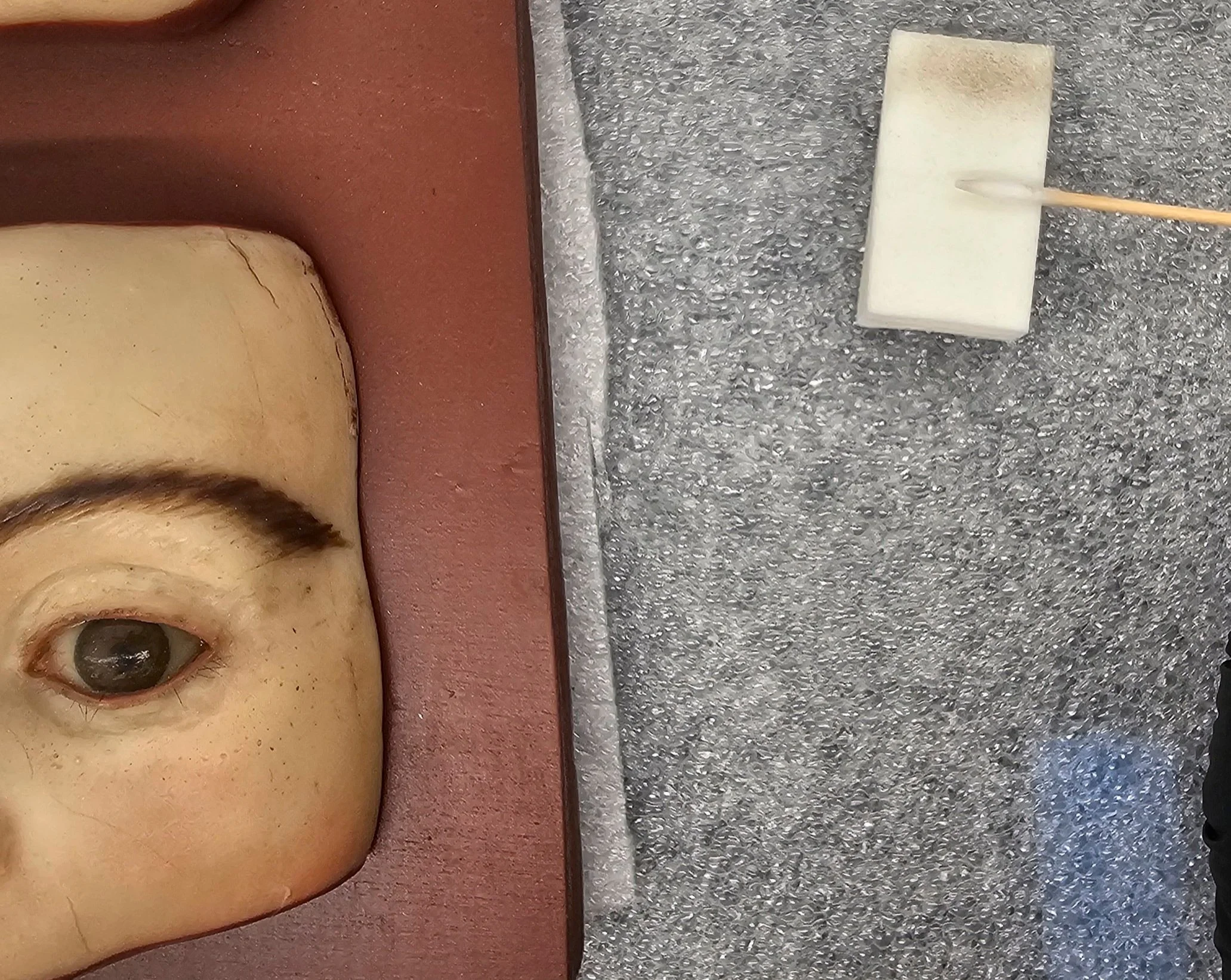
Fig. 8 Dirty cosmetic sponge and swab, during treatment
One of the difficulties of cleaning wax stems from its tendency to soften with high temperatures causing dust and debris to sink and embed over time. These embedded particles cannot be removed without overcleaning the wax. As 4 of the panels have been on continuous display for years, there is an accumulation of dust and debris across the surface and embedded in the wax, while the panel in storage, that is, without the heat of exhibition lighting, had a thick layer of dust but a lesser amount of embedded material.
After evaluating a variety of cleaning methodologies, the system we decided on is thorough dry cleaning with cosmetic sponges followed by a customized aqueous solution.
The solution applied using swabs and small soft-bristled brushes effectively cleans most of the grime off the surface without damaging the wax. For stubborn areas of grime, tissue was used for a longer dwell time and to help pull the dirt out of pits and crevices.
Vid. 1 cornea cleaning
Cornea repair
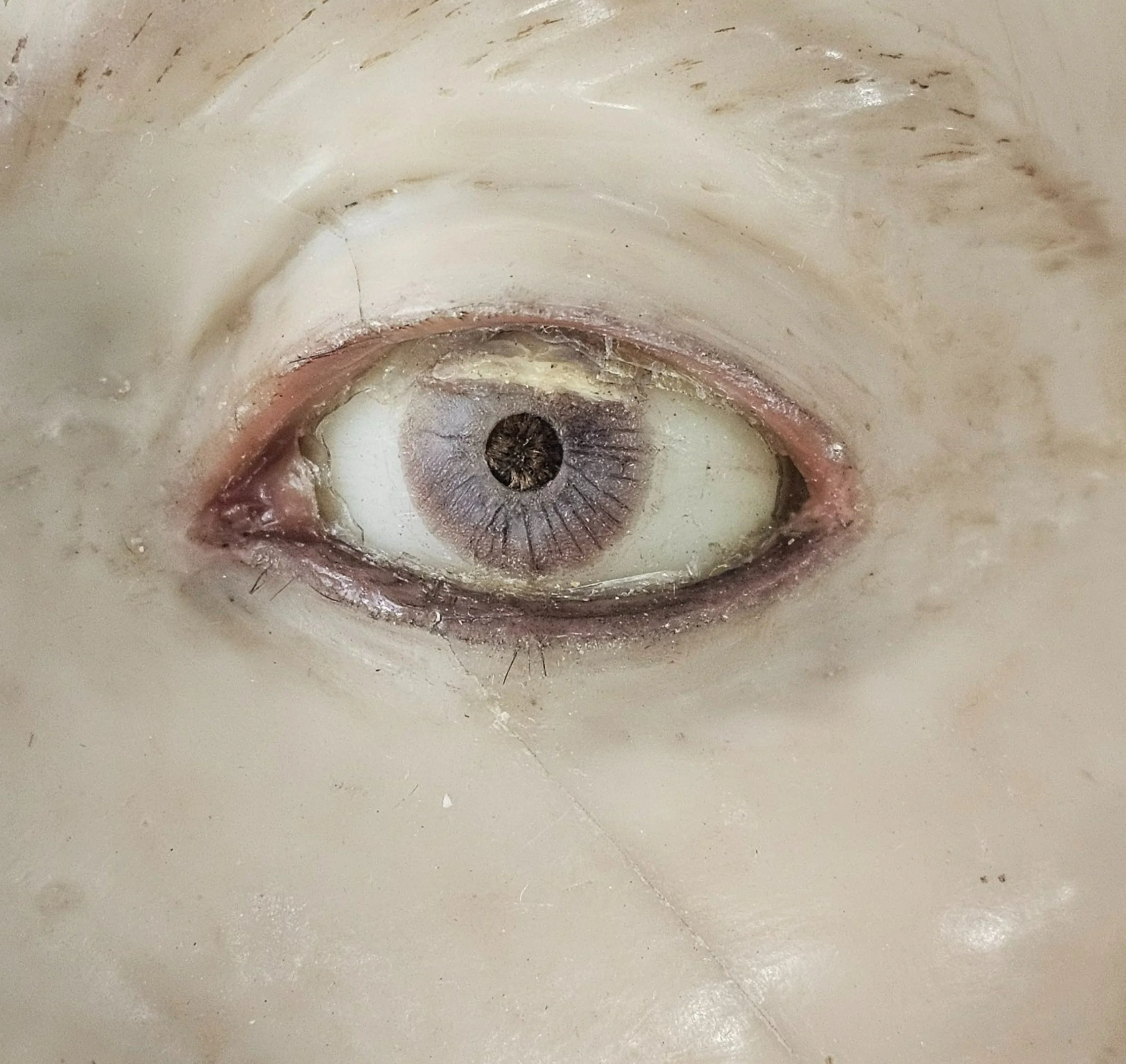
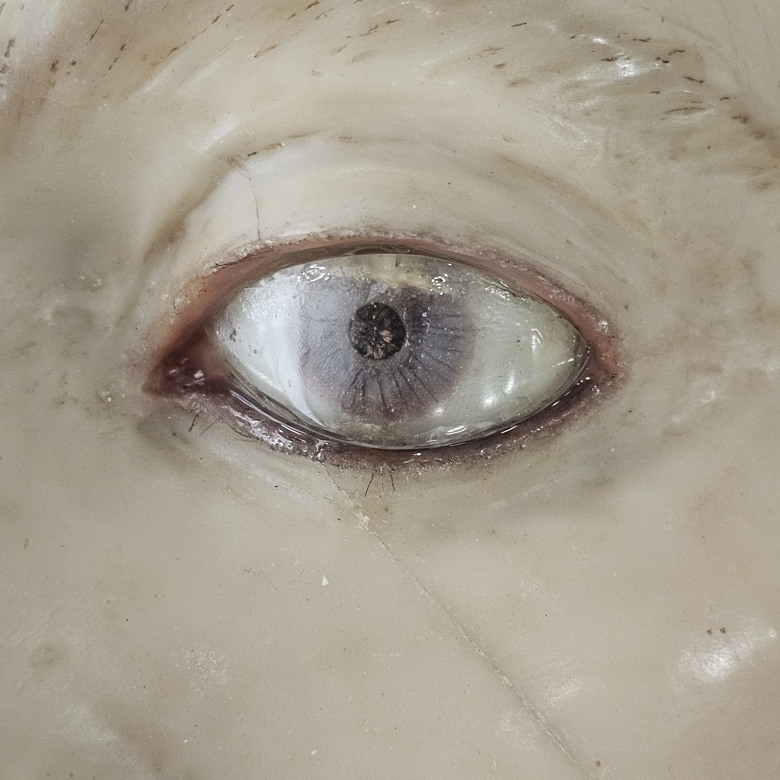
Some of the eyes exhibited cracks, distortion and cloudy discoloration of the cornea layer, which distracted from the pathology meant to be shown by the moulage. Examination of these eyes revealed two layers of material on top of a glass eye - the lower layer on top of the glass appearing to be a gelatin substance followed by a harder plasticky-coating, perhaps a later attempt at consolidation of the split gelatin cornea.
Testing found that in some cases the cracks could be reconstituted with brush applied isopropanol (see video 1). In a few severe cases the best way to approach the degraded corneal film was to remove it completely. (see fig. 9a-e).
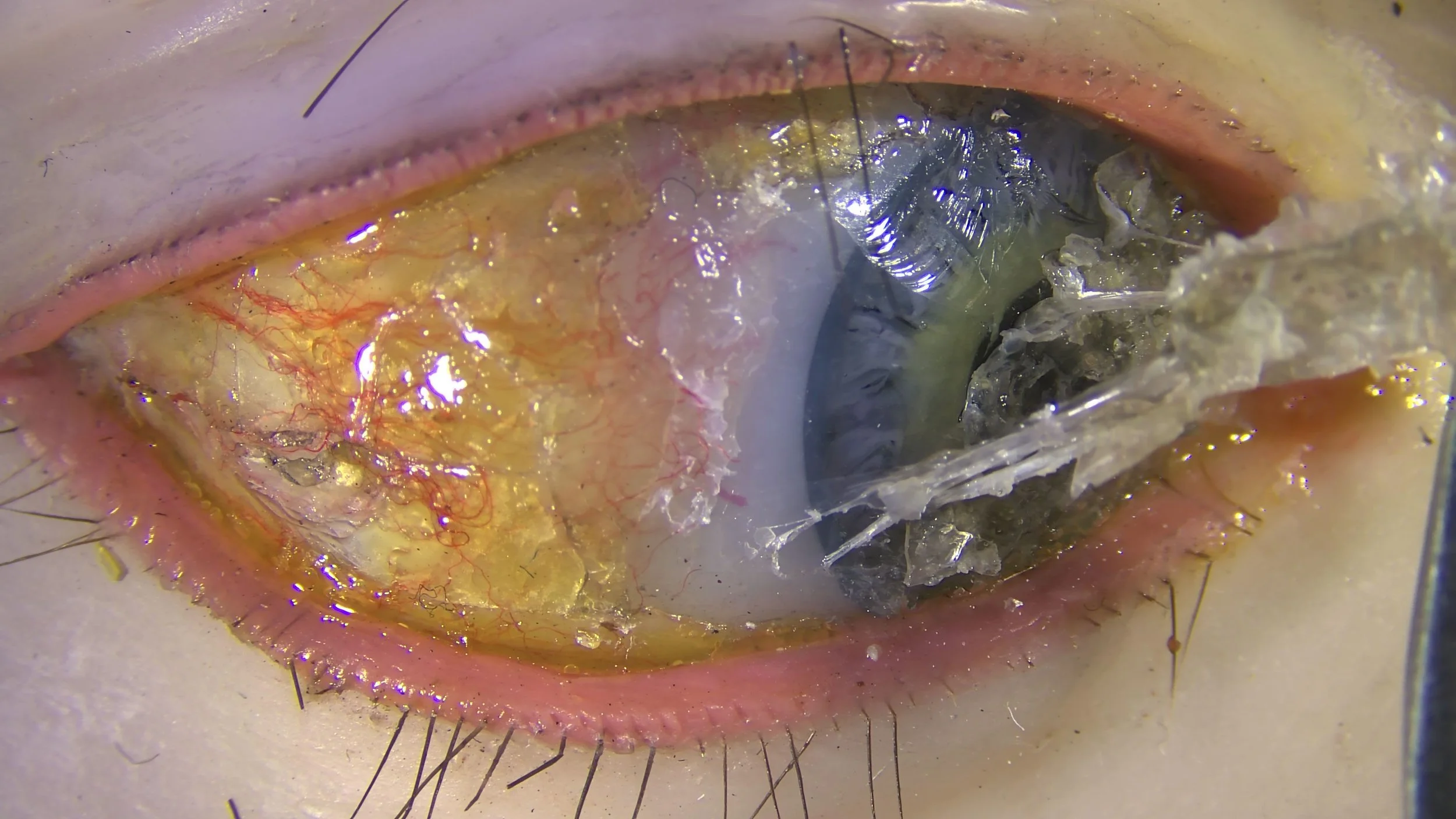
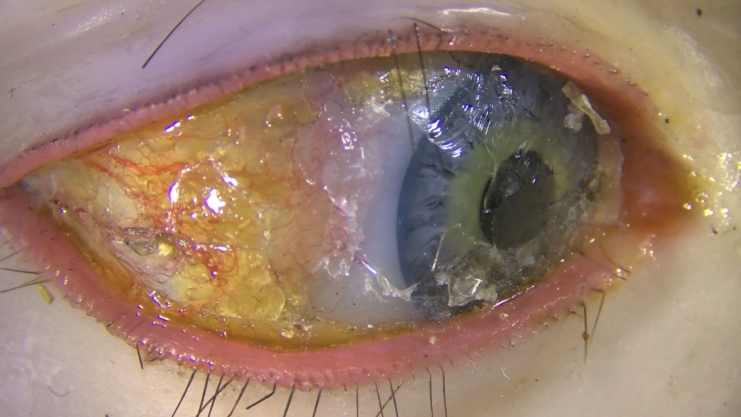
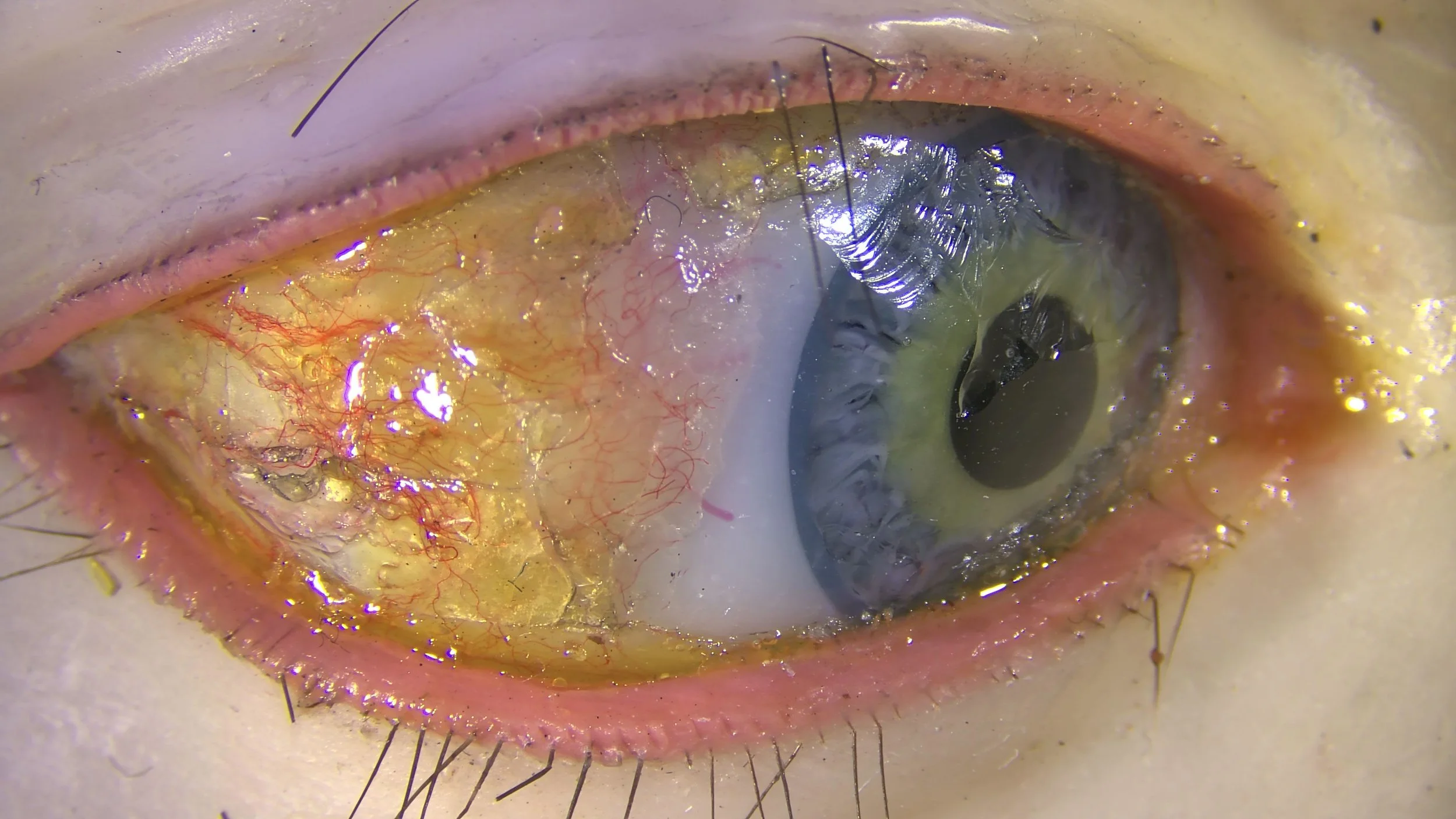
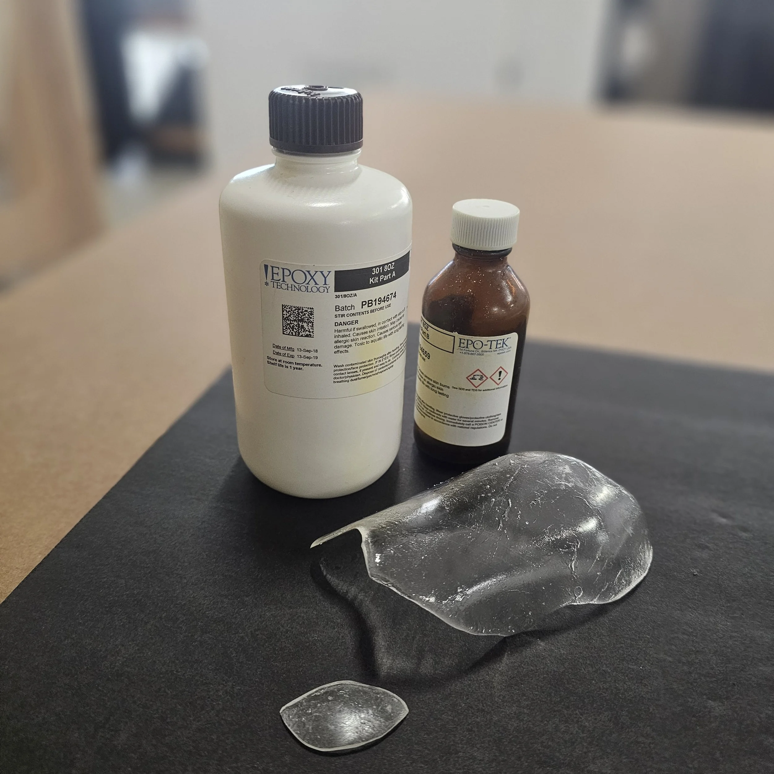
In one case the cornea had shattered and shriveled, and upon removal revealed not a glass sclera and iris, but a wax one. For this eye the original shell was removed, a cast taken of the eye, and a new shell was created with clear epoxy to protect the now exposed wax cornea (fig. 10a-d).
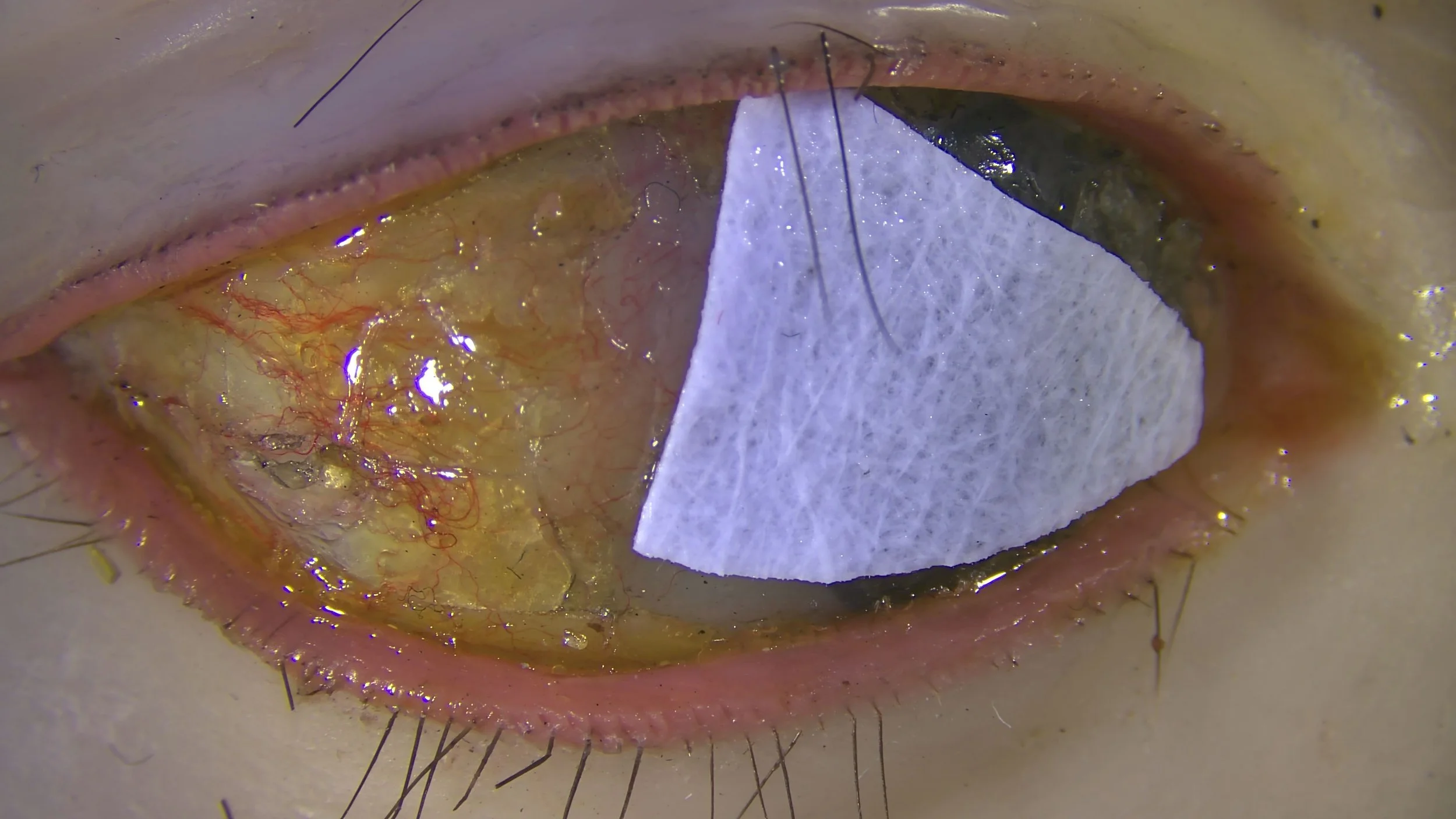
Vid. 2. Removing the degraded corneal films.
Vid. 3 applying synthetic eyelashes
Replacing the eyelashes
The original eyelashes, likely the cut ends of human hair, were embedded into the wax while it was still soft. Over time, many eyelashes were lost. Some of them fell out completely while others broke at the attachment point leaving bits of the hair embedded in the wax.
To approach the addition of new eyelashes, we created a mock-up with beeswax and synthetic lashes. This allowed us to test different adhesives and application methods before continuing treatment on the Mütter Museum eyes. Luckily we found an adhesive with ideal working properties and an application method early in our tests.
Navigating previous restoration
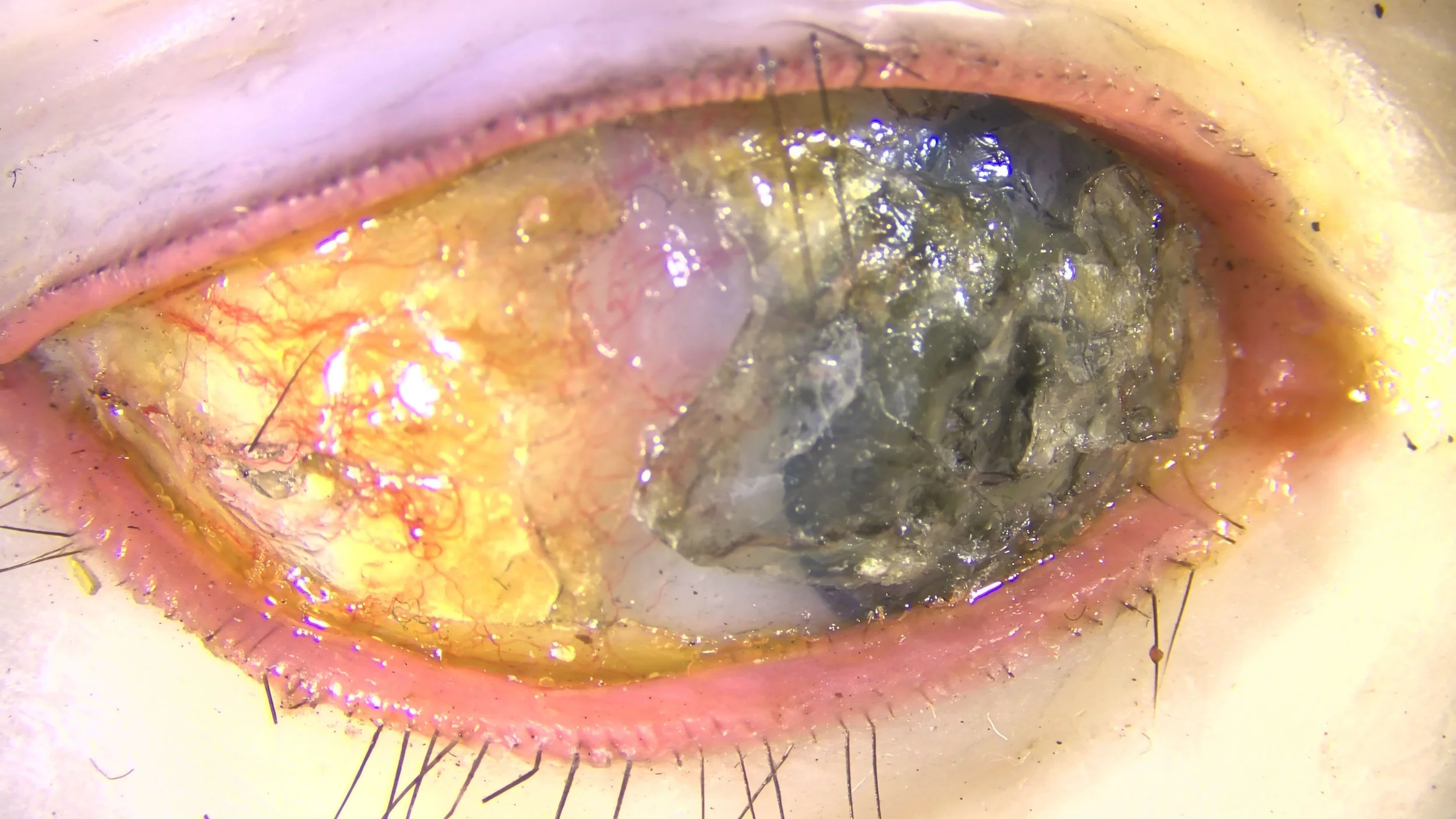
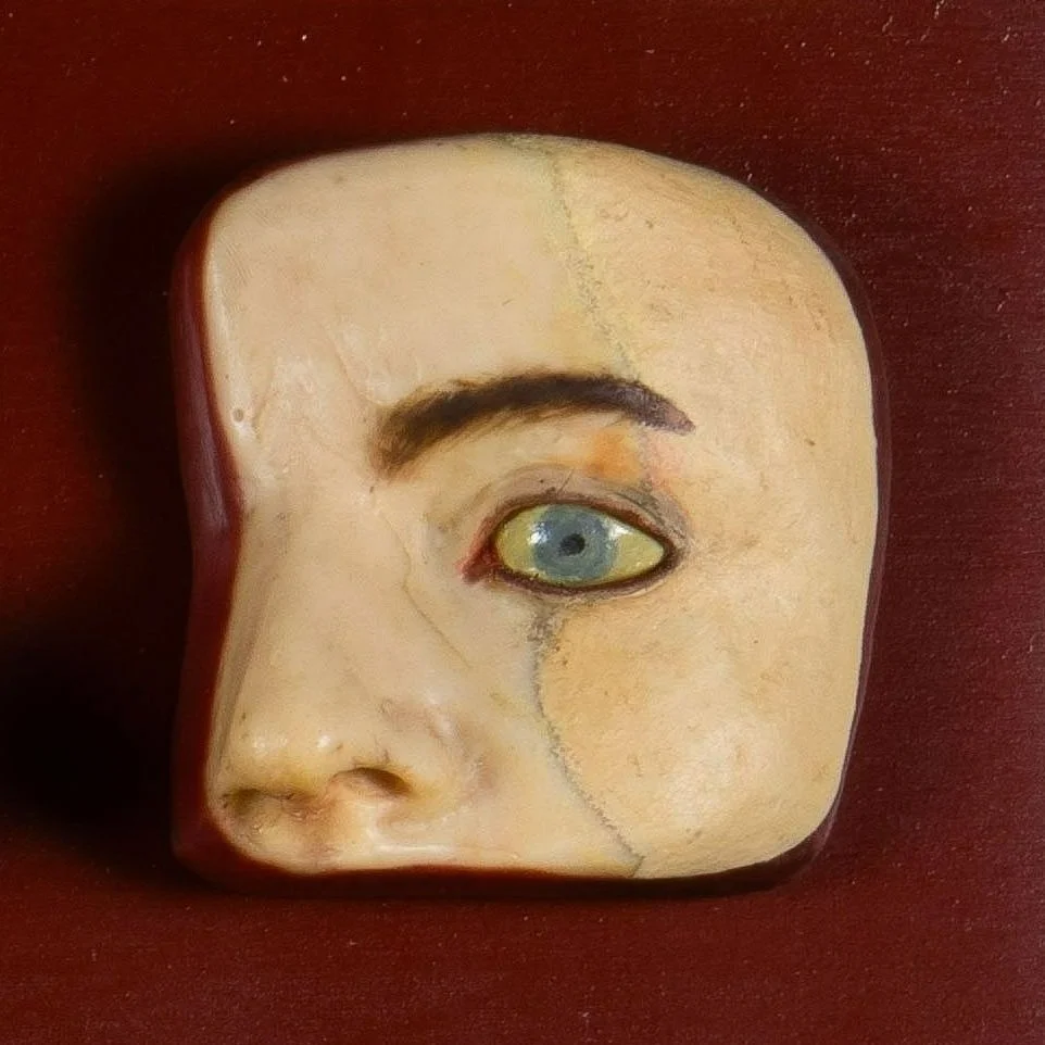
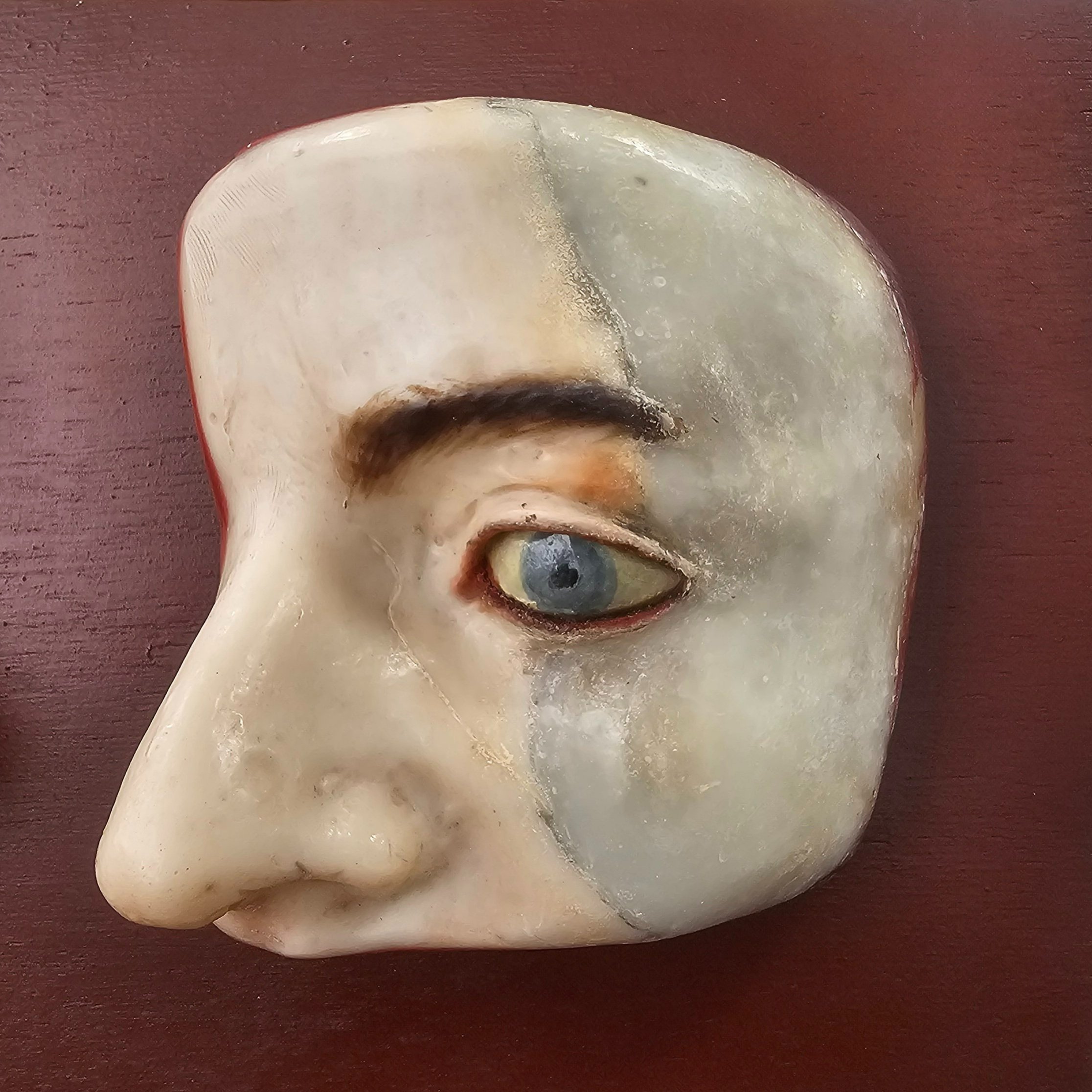
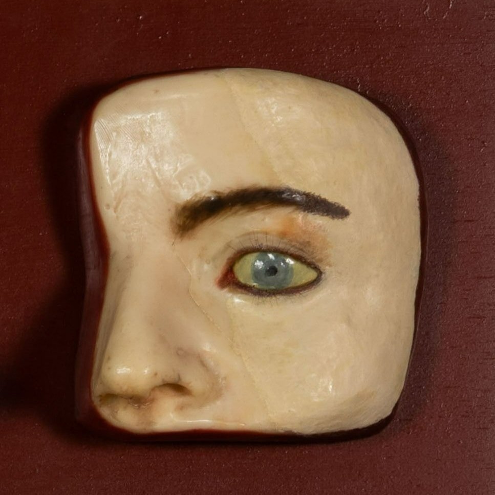
There were 3 cases of previous interventions done after the panel had suffered a fall. In these instances the restorations did not match the color or opacity of the original wax and cornea. As you can see in the before treatment photo below there was a harsh dark line between the original wax and the restoration. The overpainted cornea was opaque and flat, making it standout when compared to the depth, subtle layering, and details of the original corneas (fig. 2 and fig. 3)
Navigating a previous restoration is always a difficult task, in this case the restoration included a wax fill that was painted over. The goal was to create a smoother transition between original and added material, this was made difficult by the opacity and tone of the wax fill. by finding the right inpainting media and finding a balance with all the factors it was possible to achieve our goal.
Next time you are in Philadelphia visit the Mütter Museum and see these eyes with your own eyes!
To read more about this fascinating part of history and ceroplastics check out these resources: